DIAGNOSTICS
What is an EEG
What is an EEG (electroencephalography) test?
An electroencephalogram (EEG) is a test that measures brain activity - an extremely important diagnostic tool used in medicine. During this painless assessment, small sensor-electrodes are attached to the scalp to pick up the electrical signals produced by the brain. The brain cells communicate by means of electrical impulses and are active at all times, even during sleep. This activity appears as waves on the EEG recording. Brain waves (synchronised electrical impulses) reflect communication between areas of the brain. Brain waves can reveal information about physiological state and cognitive functioning, including stress levels or attention. These signals are recorded by a machine and analysed by a specialist. The EEG is one of the main diagnostic tests for epilepsy. It also plays a role in the diagnosis of many other disorders.
Indications for an EEG: why is an eeg test done?
An EEG test can help to diagnose and monitor a range of neurological conditions. It is used to identify the cause of some symptoms - such as memory problems - or to find out more about a condition that has already been diagnosed. An EEG test can help your physician to determine the type of epilepsy you suffer from, what may cause epileptic seizures and how most effectively to treat you. An EEG test helps doctors to assess disorders of consciousness. It can also be used additionally in the diagnosis of psychiatric and other neurological disorders.
EEG is also used in disorders such as:
- dementia
- head injury and concussion
- brain tumours
- encephalitis
- sleep disorders such as sleep apnoea.
EEG testing in children
EEG testing can also be performed in children. EEG tests are usually performed when children show developmental delays or symptoms such as loss of consciousness, headaches. The EEG will help to determine whether epileptic seizures or other neurological disorders are the cause of the symptoms.
The EEG procedure: What the EEG test looks like?
During an EEG test, the patient sits or lies down and electrodes are placed on the scalp using a special gel. The patient is asked to relax or perform specific tasks (e.g. closing and opening the eyes, deep breathing) while data are recorded on a computer for a set period of time.
Below we have described in detail how the EEG testing routine is carried out:
- You will be asked to relax in a chair or lie down on a bed.
- Electrodes will be attached to your scalp using a special paste, or a cap containing electrodes to which conductive gel is applied will be used.
- You will be asked to close your eyes, relax.
- Once the recording has started, you will be asked to remain relatively still throughout the test. You may be monitored by the technician performing the test to watch for movements that may cause possible interference with the recording, such as swallowing or blinking. The examination can be paused to allow you to rest or change position, if necessary.
- After the initial recording has been made, the specialist may test you with various stimuli. For example, you may be asked to breathe deeply and quickly for 3 minutes or you may be exposed to a bright flashing light.
- If you are being assessed for a sleep disorder, the EEG should be performed while you are asleep.
What happens after an EEG test is performed?
After the end of the EEG test, the electrodes will be removed and the paste/gel will be washed off with warm water. In some cases it may be necessary to wash the hair again at home. There may be a slight redness of the skin at the electrode sites, but this will subside within a few hours.
You will usually receive the results within 3-4 days). The EEG signal will first need to be analysed by a specialist. Our specialist can give you additional instructions after the procedure, depending on your specific situation.
EEG recording: What does a typical EEG recording look like?
Typical EEG recording is characterised by regular and symmetrical brain wave patterns. Brain waves have their own specific parameters, such as frequency and amplitude, which are taken into account when interpreting the results. These parameters, in combination with other information such as the patient's age, alertness or sleep state and location of the electrode on the scalp, are interpreted by the specialist.
The most widely known classification uses the frequency of EEG waves (alpha, beta, theta and delta)
Alpha waves - 8-13 Hz
Alpha waves occur rhythmically on both sides of the head, but often have a slightly greater amplitude on the right side, especially in right-handed people. It is a dominant rhythm in the central and occipital regions (at the back of the head), particularly prominent with eyes closed and in a relaxed state. Alpha activity tends to disappear with attention (e.g. calculations in memory, stress, or opening the eyes).
Beta waves - over 13 Hz
Beta waves tend to be small in amplitude, are usually symmetrical and more prominent at the front. Drugs such as barbiturates and benzodiazepines can increase beta waves.
Theta waves - 3.5-7.5 Hz
Theta and delta waves are collectively known as slow waves, which have a greater amplitude than faster (higher frequency) waves. Theta waves usually occur during sleep regardless of age. In conscious adults, these waves are abnormal when present in excess.
Delta waves - 3 Hz or below
Usually observed during deep sleep in adults, as well as in infants and children.
Delta waves are abnormal in a conscious adult. Delta waves can be focal (localised pathology) or diffuse (generalised dysfunction).
Other parameters assessed in the EEG recording are the morphology (i.e. shape) of the brain waves
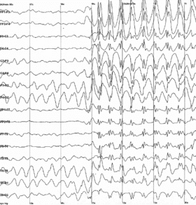
Spiking, sharp waves may be indicative of pathology, e.g. epilepsy.
Interpretation of electroencephalography results
The interpretation of the EEG results is made by a specialist who analyses the brain wave patterns and can determine the presence of abnormalities in the electrical activity of the brain. Based on these results, the clinician can make a diagnosis and recommend appropriate treatment. Clinical symptoms combined with the interpretation of the EEG results are important in making an appropriate diagnosis.


